Home » Uncategories » Compact Bone Diagram : Definition Of Bone Marrow Nci Dictionary Of Cancer Terms National Cancer Institute - Learn vocabulary, terms, and more with flashcards, games, and other study tools.
Compact Bone Diagram : Definition Of Bone Marrow Nci Dictionary Of Cancer Terms National Cancer Institute - Learn vocabulary, terms, and more with flashcards, games, and other study tools.
ads/wkwkland.txt
Compact Bone Diagram : Definition Of Bone Marrow Nci Dictionary Of Cancer Terms National Cancer Institute - Learn vocabulary, terms, and more with flashcards, games, and other study tools.. It is also called osseous tissue or cortical bone and it provides structure and support for an organism as part of its skeleton, in addition to being a location for the storage of minerals like calcium.about 80% of the weight of the human skeleton comes from. Flat bones, like those of the cranium, consist of a layer of diploë (spongy bone), lined on either side by a layer of compact bone (). Between the rings of matrix, the bone cells (osteocytes) are located in spaces called lacunae. Although the calls are close together, this type of bone is not completely solid. 0 0000 a shoutout is a way of letting people know of a.
It is also called osseous tissue or cortical bone and it provides structure and support for an organism as part of its skeleton, in addition to being a location for the storage of minerals like calcium.about 80% of the weight of the human skeleton comes from. Its repeated pattern is arranged in concentric layers of solid bone tissue. The compact bone is composed of calcified extracellular material the bone matrix and 3 major cell types which are osteoblast which ssynthesize and secrete the organic components of bone matrix which include type 1 collagen fibers proteoglycans and several glycoproteins such as ostepnectin. Apr 28, 2017 · as seen in the image below, compact bone forms the cortex, or hard outer shell of most bones in the body. If the outer layer of a cranial bone fractures, the brain is still protected by the intact inner layer.
Cartilage Bone Ossification The Histology Guide from www.histology.leeds.ac.uk The remainder is cancellous bone, which has a spongelike appearance with numerous large spaces and is found in the. (b) in this micrograph of the osteon, you can clearly see the concentric lamellae and central canals. Compact bone, as opposed to spongy bone, is made of cylindrical units, called osteons, that are tightly formed together. It is also called osseous tissue or cortical bone and it provides structure and support for an organism as part of its skeleton, in addition to being a location for the storage of minerals like calcium.about 80% of the weight of the human skeleton comes from. About press copyright contact us creators advertise developers terms privacy policy & safety how youtube works test new features press copyright contact us creators. Flat bones, like those of the cranium, consist of a layer of diploë (spongy bone), lined on either side by a layer of compact bone (). Compact bone is the strongest form of bone tissue containing few spaces. Compact bone is formed from a number of osteons, which are circular units of bone material and blood vessels.
Diagram of a typical long bone showing both cortical (compact) and cancellous (spongy) bone.
Haversian canals (sometimes canals of havers) are a series of microscopic tubes in the outermost region of bone called cortical bone. Compact bone is the denser, stronger of the two types of osseous tissue (figure 6.3.6). Compact bone is the denser stronger of the two types of bone tissue. Compact bone, also called cortical bone, dense bone in which the bony matrix is solidly filled with organic ground substance and inorganic salts, leaving only tiny spaces (lacunae) that contain the osteocytes, or bone cells.compact bone makes up 80 percent of the human skeleton; Cortical bone is compact bone while cancellous bone is trabecular and spongy bone. Microscopic structures of compact bone wedge of bone duration. A diagram of the anatomy of a bone, showing the compact bone. Between the rings of matrix the bone cells osteocytes are located in spaces called lacunae. Its repeated pattern is arranged in concentric layers of solid bone tissue. As seen in the image below, compact bone forms the cortex, or hard outer shell of most bones in the body. About press copyright contact us creators advertise developers terms privacy policy & safety how youtube works test new features press copyright contact us creators. There are pores and spaces even in compact bone. 13 photos of the compact bone diagram labeled.
The cells of compact bone, which is also called cortical bone, appear to be tightly packed into a solid mass. The two main structural components typically include spongy bone on the interior, with an outer layer of compact bone. Haversian canals (sometimes canals of havers) are a series of microscopic tubes in the outermost region of bone called cortical bone. Flat bones, like those of the cranium, consist of a layer of diploë (spongy bone), lined on either side by a layer of compact bone (). There are small canals that run through the bone, which allow blood vessels to penetrate it.
Draw The Given Diagram And Label The Following Parts A Spongy Boneb Periosteumc Yellow Marrowd Compact Bone from haygot.s3.amazonaws.com Compact bone diagram bone cross section diagram file624 diagram of compact bone new. Some, mostly older, compact bone is remodelled to form these haversian systems (or osteons). Because of its strength, the compact bone makes it possible for the bone to support weight. (b) in this micrograph of the osteon, you can clearly see the concentric lamellae and central canals. Its repeated pattern is arranged in concentric layers of solid bone tissue. Between the rings of matrix, the bone cells (osteocytes) are located in spaces called lacunae. There are pores and spaces even in compact bone. The cells of compact bone, which is also called cortical bone, appear to be tightly packed into a solid mass.
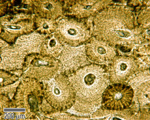
(b) in this micrograph of the osteon, you can clearly see the concentric lamellae and central canals.
Cortical bone is compact bone while cancellous bone is trabecular and spongy bone. Haversian canals (sometimes canals of havers) are a series of microscopic tubes in the outermost region of bone called cortical bone. The cells of compact bone, which is also called cortical bone, appear to be tightly packed into a solid mass. They allow blood vessels and nerves to travel through them to supply the osteocytes. Compact bone is formed from a number of osteons, which are circular units of bone material and blood vessels. Under periosteum of all bones is the bulk of the diaphysis of long bones. Compact and spongy.the names imply that the two types differ in density, or how tightly the tissue is packed together. About press copyright contact us creators advertise developers terms privacy policy & safety how youtube works test new features press copyright contact us creators. Apr 28, 2017 · as seen in the image below, compact bone forms the cortex, or hard outer shell of most bones in the body. Although the calls are close together, this type of bone is not completely solid. Diagram of a typical long bone showing both cortical (compact) and cancellous (spongy) bone. The diagram above shows a longitudinal view of an osteon. (b) in this micrograph of the osteon, you can clearly see the concentric lamellae and central canals.
Compact bone, also called cortical bone, is the hard, stiff, smooth, thin, white bone tissue that surrounds all bones in the human body. The compact bone is a dense bone found in the diaphysis. Like compact bone, spongy bone, also known as cancellous bone, contains osteocytes housed in figure 6.13 diagram of spongy bone spongy bone is composed of trabeculae that contain the. Andrew kirmayer a diagram of the anatomy of a bone, showing the compact bone. About press copyright contact us creators advertise developers terms privacy policy & safety how youtube works test new features press copyright contact us creators.
Compact And Spongy Bone Microanatomy Of Bone Tissue Bone Tissue Anatomy Bones Skeletal System Anatomy Bones from i.pinimg.com (b) in this micrograph of the osteon, you can clearly see the concentric lamellae and central canals. The remainder is cancellous bone, which has a spongelike appearance with numerous large spaces and is found in the. There are pores and spaces even in compact bone. Compact and spongy.the names imply that the two types differ in density, or how tightly the tissue is packed together. The compact bone is composed of calcified extracellular material the bone matrix and 3 major cell types which are osteoblast which ssynthesize and secrete the organic components of bone matrix which include type 1 collagen fibers proteoglycans and several glycoproteins such as ostepnectin. As seen in the image below, compact bone forms the cortex, or hard outer shell of most bones in the body. To recognise bone and understand its structure and to understand the processes by which bone can be formed. Because of its strength, the compact bone makes it possible for the bone to support weight.
(b) in this micrograph of the osteon, you can clearly see the concentric lamellae and central canals.
They allow blood vessels and nerves to travel through them to supply the osteocytes. Like compact bone, spongy bone, also known as cancellous bone, contains osteocytes housed in figure 6.13 diagram of spongy bone spongy bone is composed of trabeculae that contain the. Compact bone diagram osteon compact bone ap pinterest anatomy human anatomy and. The compact bone can be seen as the layer just underneath the periosteum, color both ends. Haversian canals (sometimes canals of havers) are a series of microscopic tubes in the outermost region of bone called cortical bone. The compact bone is a dense bone found in the diaphysis. Compact bone, also called cortical bone, is the hard, stiff, smooth, thin, white bone tissue that surrounds all bones in the human body. Andrew kirmayer a diagram of the anatomy of a bone, showing the compact bone. There are two types of bone tissue: There are small canals that run through the bone, which allow blood vessels to penetrate it. The two main structural components typically include spongy bone on the interior, with an outer layer of compact bone. Compact and spongy.the names imply that the two types differ in density, or how tightly the tissue is packed together. Compact bone diagram bone cross section diagram file624 diagram of compact bone new.
ads/wkwkland.txt
0 Response to "Compact Bone Diagram : Definition Of Bone Marrow Nci Dictionary Of Cancer Terms National Cancer Institute - Learn vocabulary, terms, and more with flashcards, games, and other study tools."


0 Response to "Compact Bone Diagram : Definition Of Bone Marrow Nci Dictionary Of Cancer Terms National Cancer Institute - Learn vocabulary, terms, and more with flashcards, games, and other study tools."
Posting Komentar